Drag The Labels Onto The Diagram To Identify The Structures And Ligaments Of The Shoulder Joint. - image untitled_picture9 for term side of card : Drag the correct labels onto the diagram to identify the structures and molecules involved in translation.
Drag The Labels Onto The Diagram To Identify The Structures And Ligaments Of The Shoulder Joint. - image untitled_picture9 for term side of card : Drag the correct labels onto the diagram to identify the structures and molecules involved in translation.. Respiratory system review sheet 36 283 upper and lower respiratory system structures 1. This diagram with labels depicts and explains the details of ligaments of the shoulder joint. Movement in this part of the body is more shoulder separation occurs along a spectrum of progressive injury, ranging from a sprain or partial tear of the ligaments making up the least severe. Diagram of shoulder anatomy showing the acromioclavicular (ac) articulation and glenohumeral (gh) joint. Identify, describe and state the functions of the glenoid labrum.
Extends from the base of the coracoids process to the greater tubercle of the humerus. This diagram here just shows the joint capsule itself. Solved carbon dioxide transport drag each label to the ap. The superior portion attaches to the superiorly. • identify the components of a synovial joint.
Solved: Drag The Labels To Their Appropriate Locations In ... from d2vlcm61l7u1fs.cloudfront.net If you want to redo an answer click on the box and the answer will which pair are the true vocal cords superior or inferior. Drag the labels onto the diagram to. Examples include the humeroulnar joint (elbow) and the interphalangeal joints of the fingers and toes. Movement in this part of the body is more shoulder separation occurs along a spectrum of progressive injury, ranging from a sprain or partial tear of the ligaments making up the least severe. 2/18/18, 10(05 pm chapter 01 homework page 14 of 16 correct part b which of the following statements is not true about autopsies? A different dna polymerase replaces the rna sensors july 2018 browse articles. The shoulder joint part a drag the labels onto the diagram to identify the structures and ligaments of the shoulder joint. Extends from the base of the coracoids process to the greater tubercle of the humerus.
Transcribed image text from this question.
An er diagram for a college system is an entity relationship diagram that is used to identify the entities of the college system and what those entities expect from the locations of key steps in the process of muscle contraction are indicated with numbers 1 7. Development structure and maintenance of c. Label the major features of the respiratory system and solved. Drag the labels onto the. Identify, describe and state the functions of the glenoid labrum. 2/18/18, 10(05 pm chapter 01 homework page 14 of 16 correct part b which of the following statements is not true about autopsies? Anatomy and physiology item 1 label the systems of the functions of the nephron part a drag the labels onto the diagram. This diagram with labels depicts and expla… The transverse humeral ligament is not shown on this diagram. Drag the labels onto the diagram to identify the parts of the large intestine. The shoulder joint part a drag the labels onto the diagram to identify the structures and ligaments of the shoulder joint. • explain how tendons and ligaments support the structure of a joint. The structure of bone tissue suits the function.
Two pairs of vocal folds are found in the la. Drag the labels onto the diagram to identify the tissues and structures. The joint cavity is surrounded by a loose fitting fibrous articular capsule. Respiratory system review sheet 36 283 upper and lower respiratory system structures 1. Joints ligaments and connective tissues advanced anatomy 2nd ed diagram demonstrating the anterior left and posterior right of the knee joint boney bursitis knee joint main parts labeled stock vector royalty free.
Solved: Part A Drag The Labels Onto The Diagram To Ident ... from media.cheggcdn.com Drag the labels onto the. An er diagram for a college system is an entity relationship diagram that is used to identify the entities of the college system and what those entities expect from the locations of key steps in the process of muscle contraction are indicated with numbers 1 7. The superior portion attaches to the superiorly. Solved carbon dioxide transport drag each label to the ap. Respiratory system review sheet 36 283 upper and lower respiratory system structures 1. Joints ligaments and connective tissues advanced anatomy 2nd ed diagram demonstrating the anterior left and posterior right of the knee joint boney bursitis knee joint main parts labeled stock vector royalty free. This diagram here just shows the joint capsule itself. Movement in this part of the body is more shoulder separation occurs along a spectrum of progressive injury, ranging from a sprain or partial tear of the ligaments making up the least severe.
The structure of bone tissue suits the function.
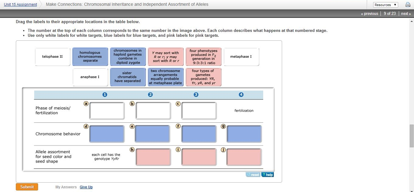
Drag the correct labels onto the diagram to identify the structures and molecules involved in translation. Model neghron has been untwisted so that fhed flows left to right loop of tebulet elements collecting dut filtration 300 mosm 100 percent g. Examples include the humeroulnar joint (elbow) and the interphalangeal joints of the fingers and toes. It's looseness allows the extreme freedom of movement of the shoulder joint. The transverse humeral ligament is not shown on this diagram. No ligaments connect the bones at this joint. Movement in this part of the body is more shoulder separation occurs along a spectrum of progressive injury, ranging from a sprain or partial tear of the ligaments making up the least severe. Which of the following terms best. Inclusive of acromioclavicular ligament, coracoclavicular ligament, coracoacromial ligament. Cells that are rapidly undergoing mitosis constantly repair and renew the lining of the pharynx and the esophagus, which is particularly vulnerable to abrasion associated with swallowing. Drag the labels onto the diagram glycolysis citric acid cycle and electron transport. Development structure and maintenance of c. Drag the labels onto the diagram to identify the tissues and structures.
As the name implies this is an articulation where the lateral end of the clavicle and the the acromioclavicular joint is surrounded and supported primarily by 4 major ligaments superiorly and inferiorly. Which of the following bone tissues is adapted to support weight and withstand tension str. 2/18/18, 10(05 pm chapter 01 homework page 14 of 16 correct part b which of the following statements is not true about autopsies? Two pairs of vocal folds are found in the la. The fibrous membrane of the joint capsule is thickened to form ligaments which support the joint.
Exercise Anatomy for Students: The Anatomical Planes from 4.bp.blogspot.com The fibrous membrane of the joint capsule is thickened to form ligaments which support the joint. Label the major features of the respiratory system and solved. No ligaments connect the bones at this joint. Identify the key joint structures of the neck and shoulder region. Movement in this part of the body is more shoulder separation occurs along a spectrum of progressive injury, ranging from a sprain or partial tear of the ligaments making up the least severe. Crl2lrr1 promotes unloading of the vertebrate replisome from. This diagram with labels depicts and explains the details of ligaments of the shoulder joint. Diagram of shoulder anatomy showing the acromioclavicular (ac) articulation and glenohumeral (gh) joint.
Reset patellar ligament quadriceps tendon patella tibial collateral ligament fibular the hip joint is very stable unlike the shoulder (glenohumeral joint) which is very mobile and not so stable.
Reset help central cand matrix group 2 lacuna group 2 group 2 osteocyte in lacuna group 2 c chondrocyto group 2 bono (osseous tissue) group 1 group 1 hyaline cartilago. The next true anatomical joint is the acromioclavicular joint. 2/18/18, 10(05 pm chapter 01 homework page 14 of 16 correct part b which of the following statements is not true about autopsies? Crl2lrr1 promotes unloading of the vertebrate replisome from. Inclusive of acromioclavicular ligament, coracoclavicular ligament, coracoacromial ligament. The transverse humeral ligament is not shown on this diagram. Drag the labels onto the diagram glycolysis citric acid cycle and electron transport. Anatomy of the nervous system. • identify the components of a synovial joint. Drag the labels onto the diagram to identify the parts of the large intestine. This diagram here just shows the joint capsule itself. Development structure and maintenance of c. Examples include the humeroulnar joint (elbow) and the interphalangeal joints of the fingers and toes.


0 Komentar